Last Updated: May 7, 2026 — Added: Candida auris dedicated section (CDC Urgent Threat, 2022-2025 case data, MALDI-TOF ID), new antifungals (ibrexafungerp 2021, oteseconazole 2022, rezafungin 2023), CLSI M27/M44/M60 and EUCAST EDef 7.3.2 standards named, antifungal drug class comparison table, MFC vs MIC, Nakaseomyces glabrata reclassification, CBPs vs ECVs, biofilm testing methods.
Introduction: Candida, Antifungal Resistance, and the Stakes of Efficacy Testing
Candida is a genus of yeasts that constitutes part of the normal human microbiome — present on skin, mucosal surfaces, and the gastrointestinal tract of most healthy individuals. Under conditions that alter immune competence or disrupt the microbial ecosystem — prolonged antibiotic therapy, immunosuppression, central venous catheter use, abdominal surgery, ICU admission, or hematologic malignancy — Candida can transition from commensal to pathogen, causing a spectrum of infections ranging from superficial mucosal disease (oral thrush, vulvovaginal candidiasis) to life-threatening invasive candidiasis and candidemia (bloodstream infection). Crude mortality rates for candidemia range from 40–50% in hospitalized patients and exceed 65% in ICU patients without prompt antifungal therapy.
Against this clinical backdrop, antifungal efficacy testing occupies a critical role in modern infectious disease management. Antifungal susceptibility data guide empirical and targeted treatment selection, detect resistance emergence before clinical failure, support surveillance of global antifungal resistance trends, and underpin the development of new antifungal drugs at a time when the pipeline has historically lagged far behind antibacterial drug development. This article covers the methods, standards, and recent clinical and scientific developments essential for laboratory professionals, clinicians, and pharmaceutical researchers engaged in antifungal efficacy testing for Candida species.
ContractLaboratory.com connects pharmaceutical companies, healthcare organizations, and research institutions with specialized microbiology and pathogen detection laboratories and antimicrobial resistance testing specialists for the full range of antifungal efficacy testing. See also our guide to fungus testing and analysis.
Candida Species and Clinical Relevance
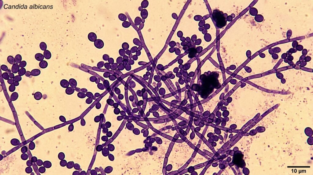
Candida albicans remains the most prevalent cause of candidiasis globally, responsible for the majority of oral, esophageal, vaginal, and systemic infections. Its virulence factors (morphological switching between yeast and hyphal forms, adhesins, secreted aspartyl proteases, biofilm formation) make it a uniquely adaptable pathogen. However, the epidemiology of invasive candidiasis has shifted substantially over the past two decades, with non-albicans species — particularly those with intrinsic or acquired resistance to first-line antifungals — accounting for an increasing proportion of infections in immunocompromised and hospitalized patients.
Clinically important Candida species and their resistance profiles:
- Candida albicans: Generally susceptible to azoles, echinocandins, and amphotericin B. Azole resistance via ERG11 mutations and efflux pump upregulation is acquired but uncommon in most settings.
- Nakaseomyces glabrata (formerly Candida glabrata): Intrinsic reduced susceptibility to azoles; echinocandin resistance via FKS1/FKS2 mutations is an increasing clinical problem. The reclassification from Candida to Nakaseomyces was established in 2020 — many clinical labs still use the old C. glabrata name.
- Candida parapsilosis: Intrinsically reduced echinocandin susceptibility (natural FKS polymorphisms). Common in neonatal and pediatric candidiasis. Azole-resistant strains are emerging in some geographic regions.
- Candida tropicalis: Important in neutropenic patients; azole resistance is increasingly documented in the Asia-Pacific. Generally susceptible to echinocandins and amphotericin B.
- Pichia kudriavzevii (formerly Candida krusei): Intrinsically resistant to fluconazole due to low affinity of ERG11 for fluconazole. Also reclassified in 2020, clinical labs widely retain the C. krusei name.
- Candida auris: See dedicated section below. CDC Urgent Threat; WHO Critical Priority pathogen. Multidrug-resistant; identification challenges; nosocomial transmission.
⚠ Candida auris: A Public Health Urgent Threat (2022–2026 Update)
Candida auris has emerged as one of the most serious global infectious disease threats of the 2020s. Originally identified in Japan in 2009 (retrospectively in South Korea as early as 1996), C. auris spread globally with unprecedented speed across geographically distinct, genetically unrelated clades — a pattern suggesting simultaneous independent emergence rather than single-source spread. In 2019, the CDC became the first public health agency to classify a fungal pathogen as an Urgent Threat — and C. auris was that pathogen. In 2022, the WHO designated C. auris as a Critical Priority human fungal pathogen, its highest tier, in the first-ever WHO Fungal Priority Pathogens List.
2022–2025 US Epidemiology
The CDC’s Antimicrobial Resistance (AR) Laboratory Network analyzed 8,033 C. auris clinical isolates from 2022 and 2023, with the following resistance findings:
- >95% resistant to fluconazole (first-line azole antifungal) — with rates exceeding 90% in all US regions
- 15% resistant to amphotericin B (liposomal and conventional formulations)
- 1% resistant to echinocandins (caspofungin, micafungin, anidulafungin) — the first-line treatment class for C. auris
- <1% pan-resistant (resistant to all three major antifungal classes) — though two echinocandin-resistant and pan-resistant C. auris outbreaks in 2023 were the first documented transmission of this resistance pattern in US healthcare settings
US case burden has expanded dramatically: from 51 clinical cases in 2016 to 4,514 in 2023 — a nearly 90-fold increase. As of 2024, C. auris infections have been reported in 39 states. This trajectory has led the CDC to maintain echinocandins as the empirical first-line treatment — pending susceptibility testing results — given their retained activity against the majority of current isolates.
Why C. auris Is Uniquely Difficult to Manage
- Identification failures: Many conventional biochemical identification systems (including the Vitek 2, API 20C AUX in pre-updated versions) misidentify C. auris as other Candida species — most commonly C. haemulonii, C. famata, or Rhodotorula glutinis. This misidentification can result in inappropriate therapy and missed infection control responses.
- MALDI-TOF MS with updated databases required: Matrix-Assisted Laser Desorption/Ionization Time-of-Flight Mass Spectrometry (MALDI-TOF MS) with current, updated spectral libraries (Bruker MALDI Biotyper or bioMérieux VITEK MS with C. auris-specific entries) is now the recommended method for definitive C. auris identification in clinical settings. Molecular sequencing (ITS region or D1/D2 domain of 28S rDNA) remains the gold standard for confirmation.
- Environmental persistence: C. auris survives on dry hospital surfaces for weeks to months — a property shared with problematic bacteria like MRSA but unusual for fungi. Standard hospital disinfectants, including many quaternary ammonium compounds, are often ineffective. EPA-registered disinfectants with demonstrated efficacy against C. auris and enhanced terminal cleaning procedures are required for outbreak control.
- Nosocomial transmission: Unlike most Candida species, C. auris spreads efficiently through healthcare settings via person-to-person transmission and environmental contamination — more analogous to MRSA or VRE transmission than to typical endogenous Candida infections.
- Testing challenges for C. auris: Established CLSI clinical breakpoints are not yet available for all drug-C. auris combinations. The CDC has published tentative breakpoints to guide clinical decision-making. Susceptibility testing for C. auris must be performed by CLSI M27 broth microdilution — commercial panels are beginning to include C. auris, but availability varies.
Antifungal Drug Classes: Mechanisms and Resistance
Understanding the mechanism of action and resistance patterns of each antifungal drug class is an essential context for interpreting efficacy testing results. Three established classes — plus two recently approved novel agents — constitute the current armamentarium against Candida:
| Drug class | Key agents | Activity type | Target / mechanism | Resistance mechanisms |
| Azoles | Fluconazole, voriconazole, itraconazole, posaconazole, isavuconazole | Fungistatic against Candida | Inhibit lanosterol 14α-demethylase (CYP51/ERG11) → block ergosterol biosynthesis | ERG11 point mutations (reduce drug binding); TAC1/MRR1-mediated upregulation of efflux pumps (CDR1/CDR2, MDR1); ERG3 loss |
| Echinocandins | Caspofungin, micafungin, anidulafungin, rezafungin (newest, 2023) | Fungicidal against most Candida | Inhibit β-1,3-D-glucan synthase (FKS1/FKS2 subunit) → disrupt cell wall integrity | FKS1 or FKS2 hot-spot mutations (reduce enzyme affinity); overexpression of chitin synthase (compensatory); C. parapsilosis natural FKS polymorphisms |
| Polyenes | Amphotericin B (conventional, liposomal, lipid complex), nystatin (topical) | Fungicidal; broad-spectrum | Bind ergosterol in fungal cell membrane → form pores → membrane depolarization and cell death | Rare ERG mutations reducing ergosterol content; biofilm-mediated tolerance; not typically transmissible resistance |
| Triterpenoids (new class) | Ibrexafungerp (Brexafemme; FDA 2021) | Fungicidal against Candida | Inhibits β-1,3-D-glucan synthase at a different binding site than echinocandins; oral bioavailability | FKS mutations confer some cross-resistance; mechanism of full ibrexafungerp resistance under study |
| Tetrazoles (new class) | Oteseconazole (Vivjoa; FDA 2022) | Fungistatic (azole mechanism) | High-affinity CYP51 inhibitor with greater selectivity for fungal vs mammalian CYP enzymes than existing azoles | ERG11 mutations (cross-resistance expected with some azole-resistant strains); limited clinical resistance data |
Antifungal Susceptibility Testing Standards: CLSI and EUCAST
Two organizations provide the reference standards for antifungal susceptibility testing of yeasts, including Candida species. Their methods differ in important technical details — primarily inoculum preparation, glucose supplementation, incubation temperature, and endpoint reading criteria — and produce different MIC values for some drug-species combinations. Both are internationally recognized and regulatory-accepted, but specific clinical breakpoints may differ between CLSI and EUCAST for the same drug-species combination.
CLSI Standards for Yeast Susceptibility Testing
- CLSI M27 — Reference Method for Broth Dilution Antifungal Susceptibility Testing of Yeasts (4th Edition, 2017): The gold standard reference method for broth microdilution testing of Candida and other yeasts. Specifies RPMI 1640 medium with L-glutamine and without bicarbonate, buffered to pH 7.0 with MOPS; fungal inoculum of 0.5–2.5 × 10³ CFU/mL; 24-hour (or 48-hour for C. parapsilosis and C. tropicalis with azoles) incubation at 35°C; round-bottomed 96-well microtiter plate format; MIC reading criteria: ≥50% growth inhibition for azoles (trailing growth phenomenon addressed with specific reading guidelines); complete inhibition for echinocandins and amphotericin B.
- CLSI M44 — Disk Diffusion Method for Antifungal Susceptibility Testing of Yeasts (3rd Edition, 2018): The CLSI standard for disk diffusion susceptibility testing of yeasts on Mueller-Hinton agar supplemented with 2% glucose and 0.5 mg/L methylene blue. Provides zone diameter breakpoints for fluconazole, voriconazole, caspofungin, micafungin, and anidulafungin against C. albicans, C. tropicalis, and C. parapsilosis. See also our guide to Mueller-Hinton agar composition and applications.
- CLSI M60 — Performance Standards for Antifungal Susceptibility Testing of Yeasts (current edition): The companion document to M27 that provides the species-specific clinical breakpoints (susceptible/susceptible-dose dependent/intermediate/resistant) and quality control MIC ranges for CLSI M27 testing. Updated periodically as new resistance data emerge. Clinical breakpoints are established for the major Candida species against azoles, echinocandins, amphotericin B, and 5-flucytosine.
EUCAST Standards
- EUCAST EDef 7.3.2 — Method for Determination of Broth Dilution MICs of Antifungal Agents for Yeasts: The EUCAST reference broth microdilution method for yeasts. Uses RPMI 1640 buffered to pH 7.0 with MOPS; inoculum of 0.5–2.5 × 10⁵ CFU/mL (higher than CLSI M27); flat-bottomed microtiter plates; 24-hour incubation at 35–37°C. The higher EUCAST inoculum produces MIC values that are typically 1–2 dilutions higher than CLSI M27 for many drug-species combinations — a key source of method-related MIC discordance.
- EUCAST Fungal Breakpoints Table (FREF): EUCAST maintains separate clinical breakpoints for fungal pathogens, including Candida species, updated annually at eucast.org. EUCAST uses a two-category system (Susceptible / Resistant) for most drug-species combinations, with an intermediate category (Susceptible, Increased Exposure) indicating that a higher dose or altered pharmacokinetic exposure may achieve efficacy.
Methods for Antifungal Efficacy Testing
1. Broth Microdilution — The Gold Standard
The broth microdilution method (CLSI M27 / EUCAST EDef 7.3.2) is the reference standard for Candida antifungal susceptibility testing. Serial two-fold dilutions of the antifungal agent are prepared in 96-well microtiter plates, each well containing the RPMI 1640 broth medium. A standardized Candida inoculum is added, and plates are incubated for 24–48 hours. The
- Minimum Inhibitory Concentration (MIC): The lowest antifungal concentration that inhibits visible fungal growth by the defined endpoint criterion (≥50% for azoles; complete inhibition for echinocandins/amphotericin B). The MIC is compared against published clinical breakpoints (CLSI M60 or EUCAST FREF) to classify the isolate as Susceptible, Susceptible-Dose Dependent, Intermediate, or Resistant.
- Minimum Fungicidal Concentration (MFC): Determined by subculturing wells showing no visible growth onto drug-free agar to assess fungal viability. The MFC is the lowest concentration that kills ≥99.9% of the initial inoculum. The MIC/MFC ratio distinguishes fungistatic agents (MFC >> MIC, e.g., azoles against Candida) from fungicidal agents (MFC ≈ MIC, e.g., echinocandins and amphotericin B against most Candida species). Fungicidal activity is particularly important for immunocompromised patients who require pathogen killing rather than growth inhibition.
Trailing growth phenomenon: A challenge unique to Candida azole testing — wells at concentrations above the MIC may show faint turbidity (trailing growth) because azoles are fungistatic, and a small population of cells continues to grow even at elevated drug concentrations. CLSI M27 addresses this with specific endpoint reading criteria (50% inhibition vs 100%), but trailing growth remains a source of inter-laboratory variability in MIC readings.
2. Disk Diffusion Method
The disk diffusion (Kirby-Bauer) method is performed on Mueller-Hinton agar supplemented with 2% glucose and methylene blue per CLSI M44. Antifungal-impregnated disks (fluconazole 25 μg; voriconazole 1 μg; caspofungin 5 μg; micafungin 10 μg; anidulafungin 10 μg) are placed on agar plates inoculated with a standardized Candida suspension. After incubation, inhibition zone diameters are measured and compared to CLSI M44 zone diameter breakpoints.
Disk diffusion is less expensive and more practical than broth microdilution for routine clinical volume, but has important limitations: it cannot be used for amphotericin B (which does not diffuse adequately in agar) or for Candida auris (zone diameter breakpoints not established). Disk diffusion is applicable only for species and drug combinations explicitly validated in CLSI M44 and EUCAST antifungal disk diffusion tables.
3. E-test (Gradient Strip Method)
The E-test (now marketed under various names, including Etest by bioMérieux) uses a plastic strip impregnated with a continuous exponential gradient of antifungal agent concentration. The strip is placed on drug-free agar inoculated with Candida, and the MIC is read at the point where the inhibition ellipse intersects the strip’s calibrated concentration scale. The E-test provides a continuous MIC estimate without requiring multi-well dilution equipment and is widely used in clinical microbiology for yeast susceptibility. Important caveats: results may differ from the reference broth microdilution MIC by ≥2 doubling dilutions for some drug-species combinations, particularly echinocandins.
4. Clinical Breakpoints vs Epidemiological Cutoff Values
Two distinct interpretive criteria are applied to antifungal MIC results, serving different purposes:
- Clinical Breakpoints (CBPs) — S/I/R categories: Established by CLSI (in M60) and EUCAST (in FREF) for specific drug-species combinations based on pharmacokinetic/pharmacodynamic (PK/PD) modeling, clinical outcome data, and MIC distribution data. CBPs define Susceptible (S), Susceptible-Dose Dependent or Intermediate (I), and Resistant (R) categories that directly guide treatment decisions. CBPs are only established where sufficient clinical and microbiological data exist — this means that for C. auris and some drug combinations, formal CLSI CBPs are not yet available. The CDC has published tentative C. auris-specific breakpoints to bridge this gap.
- Epidemiological Cutoff Values (ECVs / ECOFFs): Defined by CLSI (in M59) and EUCAST to separate the wild-type (WT) population — organisms with no acquired or mutational resistance mechanisms — from non-wild-type (NWT) strains that harbor detectable resistance. ECVs are based purely on MIC distribution analysis and do not incorporate clinical outcome data. They are valuable for resistance surveillance (detecting emerging resistance trends before clinical breakpoints have been established) and for drug-species combinations where CBPs don’t exist.
5. Flow Cytometry and Automated Systems
Flow cytometry enables rapid, objective assessment of antifungal activity by measuring cell viability, membrane integrity, and metabolic activity of Candida cells exposed to antifungal agents — using fluorescent probes that distinguish viable from dead or compromised cells. Commercial automated susceptibility testing systems (Vitek 2 YST card, Sensititre YEASTONE) use colorimetric endpoints (Alamar Blue reduction) in microtiter format to provide semi-automated MIC determination within 24 hours. Automated systems offer practical advantages for high-volume clinical laboratories but require validation of their performance against the CLSI M27 reference method for each drug-species combination, particularly for C. auris.
6. MALDI-TOF MS for Fungal Species Identification
Accurate species identification is the prerequisite for any antifungal susceptibility test, and its importance has intensified with the emergence of C. auris. Matrix-Assisted Laser Desorption/Ionization Time-of-Flight Mass Spectrometry (MALDI-TOF MS) — which produces a protein mass spectrum that serves as a unique molecular fingerprint for microbial species — is now the standard of care for rapid fungal identification in clinical microbiology, reducing identification time from 24–72 hours (conventional biochemical methods) to minutes.
For C. auris specifically, MALDI-TOF MS requires: (1) an updated spectral database including validated C. auris reference spectra; and (2) sufficient C. auris-specific entries for accurate discrimination from the closely related C. haemulonii species complex. Both Bruker Biotyper (with online database updates) and bioMérieux VITEK MS (with SARAMIS database updates) now provide reliable C. auris identification when databases are current. Molecular sequencing (ITS2 or D1/D2 of 28S rDNA) remains the gold standard for ambiguous cases.
Biofilm-Associated Candida: Special Testing Considerations
Candida species — most notably C. albicans and C. parapsilosis — form biofilms on medical devices including central venous catheters, urinary catheters, prosthetic heart valves, and orthopedic implants. Biofilm formation provides profound protection against antifungal agents through multiple mechanisms: restricted drug penetration into the biofilm matrix; metabolic dormancy of deep biofilm layers; altered cell wall composition; and the presence of persister cells. Echinocandin MICs for planktonic (free-swimming) C. albicans are dramatically lower than minimum biofilm eradication concentrations (MBECs) — biofilm cells may require drug concentrations 1,000-fold higher than planktonic MICs for eradication.
Specialized in vitro biofilm susceptibility testing methods:
- XTT reduction assay: The most widely used quantitative biofilm viability assay. XTT (2,3-bis(2-methyloxy-4-nitro-5-sulpophenyl)-2H-tetrazolium-5-carboxanilide) is reduced by metabolically active biofilm cells to a colored formazan product, measured spectrophotometrically. Provides a quantitative index of biofilm metabolic activity in the presence of antifungal agents.
- Crystal violet staining: Stains the biofilm matrix (polysaccharide and protein) non-specifically; provides a measure of total biofilm biomass rather than viability. Used for initial biofilm formation quantification and screening assays.
- MBEC (Minimum Biofilm Eradication Concentration) assay: The Calgary Biofilm Device (MBEC Assay) grows biofilm on 96-peg lids, challenges them with antifungal gradients, then measures regrowth to determine the minimum concentration that eradicates biofilm. CLSI M59 provides guidelines for biofilm susceptibility testing of yeasts.
New Antifungal Agents Approved 2021–2023
Three new antifungal agents have received FDA approval since this article’s original publication — representing the most significant pipeline additions in over a decade and providing options for infections caused by resistant or refractory Candida species:
Ibrexafungerp (Brexafemme) — FDA Approved 2021
Ibrexafungerp is the first approved member of the triterpenoid antifungal class — a structurally novel glucan synthase inhibitor that targets the same FKS enzyme as echinocandins but binds at a different site on the β-1,3-D-glucan synthase complex. This distinct binding mechanism means that ibrexafungerp retains activity against many, though not all, echinocandin-resistant strains — making it particularly relevant for the treatment of infections caused by Candida isolates with FKS hot-spot mutations. Unlike echinocandins, ibrexafungerp is available in an oral formulation (tablets), providing a convenient treatment option for ambulatory and step-down therapy. Current FDA indications: vulvovaginal candidiasis and recurrent vulvovaginal candidiasis. Clinical trials for invasive candidiasis are ongoing.
Oteseconazole (Vivjoa) — FDA Approved 2022
Oteseconazole is a novel tetrazole antifungal with a mechanism analogous to azoles (CYP51 inhibition) but with significantly greater selectivity for fungal CYP51 over mammalian CYP enzymes. This improved selectivity profile reduces the off-target hormonal side effects that limit long-term azole use (testosterone suppression, cortisol interference). Oteseconazole is FDA-approved for recurrent vulvovaginal candidiasis in non-childbearing-potential women. Because of teratogenicity concerns, it is contraindicated in women who may become pregnant — a significant prescribing restriction. Oral bioavailability and prolonged half-life allow once-weekly or less frequent maintenance dosing for prophylaxis of recurrent infection.
Rezafungin (Rezzayo) — FDA Approved 2023
Rezafungin is a next-generation echinocandin distinguished by its markedly prolonged half-life (~130 hours) compared to first-generation echinocandins (caspofungin, micafungin, anidulafungin; all ~9–24 hours). This pharmacokinetic profile allows once-weekly IV dosing rather than the daily IV administration required by first-generation echinocandins — a significant convenience and potential outpatient therapy advantage. Rezafungin is FDA-approved for candidemia and invasive candidiasis in adult patients with limited or no alternative treatment options. Early in vitro data demonstrate activity against C. auris, though like all echinocandins, it remains susceptible to FKS mutations that confer echinocandin resistance.
Challenges in Antifungal Efficacy Testing
- Species-drug specificity of breakpoints: Not all breakpoints are established for all drug-species combinations. For emerging species like C. auris and for newer antifungals, breakpoints may be incomplete or provisional. Labs must recognize when MIC data cannot be directly interpreted against established breakpoints and should report raw MIC values alongside any available tentative breakpoints.
- Method-dependent MIC discordance: CLSI M27 and EUCAST EDef 7.3.2 use different inoculum sizes (1,000-fold difference), medium preparations, and incubation conditions. MIC values from these two methods are not interchangeable for all drug-species combinations. When comparing MICs across institutions or studies, the reference method must be specified.
- Trailing growth phenomenon: Problematic for azole MIC endpoint reading — reduced by methylene blue supplementation in disk diffusion but remains a challenge in broth microdilution for some isolates.
- Heteroresistance: Some Candida populations contain subpopulations with different susceptibility profiles — strains may appear susceptible by standard MIC testing yet harbor small resistant subpopulations that emerge under antifungal pressure. Population analysis profiling (PAP) methods can detect heteroresistance but are not routinely performed in clinical settings.
- PK/PD integration: MIC values alone do not determine clinical outcome. Pharmacokinetic/pharmacodynamic (PK/PD) relationships govern efficacy: azoles follow time-dependent killing (AUC/MIC ratio drives efficacy); echinocandins follow concentration-dependent killing (Cmax/MIC and AUC/MIC ratios). PK/PD target attainment modeling — integrating patient PK parameters with MIC data — is increasingly used to optimize dosing, especially for critically ill patients with altered drug distribution and clearance.
Advancements in Antifungal Efficacy Testing
- Molecular resistance detection (PCR and sequencing): Detection of specific resistance mutations in ERG11 (azole resistance), FKS1/FKS2 (echinocandin resistance), and ERG3 directly from clinical isolates or increasingly from primary clinical specimens (blood, BAL). Molecular testing provides rapid resistance guidance without waiting for phenotypic susceptibility results — typically available in hours rather than 24–48 hours. Whole-genome sequencing (WGS) provides comprehensive resistance gene information and enables outbreak tracking.
- High-throughput screening for antifungal discovery: Library-scale screening (100,000+ compounds) against Candida panel strains in miniaturized broth microdilution or fluorescent metabolic assay formats is the front-end of the antifungal discovery pipeline, enabling identification of novel chemical scaffolds for drug development.
- Combination antifungal therapy testing: Checkerboard assays (two-dimensional MIC matrices testing all pairwise combinations of two drugs) and time-kill kinetics studies assess synergy, additivity, indifference, or antagonism between antifungal combinations. Combination therapy (e.g., echinocandin + liposomal amphotericin B for invasive C. auris) may be required for pan-resistant or recalcitrant infections.
- Rapid diagnostics for invasive candidiasis: T2Candida Panel (T2 Biosystems) — a FDA-cleared molecular diagnostic that detects C. albicans, C. tropicalis, C. parapsilosis, C. glabrata, and C. krusei directly from whole blood in ~3 hours without the need for blood culture. β-D-Glucan assays (Fungitell) detect a pan-fungal cell wall biomarker in serum. These diagnostics provide earlier infection detection but do not replace susceptibility testing.
Finding Specialized Antifungal Efficacy Testing Laboratories
Antifungal efficacy testing — particularly for Candida auris and multidrug-resistant species — requires laboratories with specialized mycological expertise, CLSI M27/M44 or EUCAST EDef 7.3.2 capability, appropriate biosafety infrastructure, and experience with specialized endpoints (MFC, biofilm assays, combination testing). For pharmaceutical antifungal drug development, GLP-compliant testing may be required. For clinical diagnostic antifungal susceptibility, CLIA-certified and CAP-accredited laboratories provide regulated testing.
ContractLaboratory.com connects pharmaceutical companies, hospitals, public health agencies, and researchers with specialized microbiology and pathogen detection laboratories for all forms of antifungal susceptibility and efficacy testing. See also our fungus testing and analysis guide and antimicrobial resistance testing resources.
Frequently Asked Questions About Antifungal Efficacy Testing for Candida
The MIC (Minimum Inhibitory Concentration) is the lowest concentration of an antifungal agent that visibly inhibits fungal growth under defined test conditions. It is the standard endpoint in broth microdilution susceptibility testing per CLSI M27 or EUCAST EDef 7.3.2. The MFC (Minimum Fungicidal Concentration) goes further — it is the lowest antifungal concentration that kills ≥99.9% of the initial fungal inoculum, determined by subculturing MIC wells onto drug-free agar and counting surviving colonies. The MIC/MFC ratio reveals whether an antifungal acts as fungistatic (inhibits growth but doesn’t kill — MFC greatly exceeds MIC; typical of azoles against Candida) or fungicidal (kills at concentrations close to the MIC — typical of echinocandins and amphotericin B against most Candida). Fungicidal activity is clinically important for immunocompromised patients — in the absence of a functional immune system, a fungistatic drug may suppress but not eliminate a Candida infection, increasing relapse risk. MFC testing is not routinely performed in clinical labs but is important for antifungal drug development and research.
Candida auris was classified by the CDC as an Urgent Threat — its highest alarm tier and the first-ever fungal pathogen to receive this designation — and by WHO as a Critical Priority pathogen in 2022. Several characteristics make it uniquely dangerous: it is multidrug-resistant, with over 95% of US clinical isolates resistant to fluconazole and about 15% resistant to amphotericin B (per the CDC’s 2022-2023 analysis of 8,033 isolates), leaving echinocandins as the primary treatment option for most infections. US cases rose from just 51 in 2016 to 4,514 in 2023, now reported in 39 states. It spreads efficiently through healthcare settings via surfaces and person-to-person contact — more like MRSA than a typical Candida infection — and persists on hospital surfaces for weeks, where many standard disinfectants are ineffective. It is difficult to identify with conventional laboratory methods — many systems misidentify it as other species — requiring MALDI-TOF MS with updated databases or molecular sequencing. The emergence of pan-resistant C. auris outbreaks in 2023 represents an alarming escalation, as no established antifungal would be effective against these isolates.
CLSI (Clinical and Laboratory Standards Institute) has published three main standards for Candida antifungal susceptibility testing. CLSI M27 (Reference Method for Broth Dilution Antifungal Susceptibility Testing of Yeasts, 4th Edition) is the gold standard reference method — it specifies all technical parameters of the broth microdilution method including medium composition (RPMI 1640 with MOPS buffer), inoculum size, incubation conditions, and endpoint reading criteria. CLSI M44 (Disk Diffusion Method for Antifungal Susceptibility Testing of Yeasts, 3rd Edition) provides standardized disk diffusion methodology on glucose-supplemented Mueller-Hinton agar for routine clinical testing. CLSI M60 (Performance Standards for Antifungal Susceptibility Testing of Yeasts) contains the actual clinical breakpoints (susceptible, susceptible-dose dependent, intermediate, resistant) and quality control MIC ranges for M27 testing — this is the companion interpretive document that tells labs whether a given MIC indicates clinical susceptibility or resistance. EUCAST provides the complementary European standards (EDef 7.3.2 for broth microdilution; annual FREF breakpoint tables) with some technical differences from CLSI that can produce different MIC values for the same isolate.
Three significant new antifungals received FDA approval between 2021 and 2023. Ibrexafungerp (Brexafemme; 2021) is the first member of the triterpenoid antifungal class — an oral glucan synthase inhibitor that targets the FKS enzyme at a different binding site than echinocandins, giving it activity against some echinocandin-resistant strains. It is approved for vulvovaginal candidiasis and recurrent vulvovaginal candidiasis. Oteseconazole (Vivjoa; 2022) is a novel tetrazole with an azole-like mechanism (CYP51 inhibition) but greater selectivity for fungal over mammalian CYP enzymes; approved for recurrent vulvovaginal candidiasis in non-childbearing-potential women. Rezafungin (Rezzayo; 2023) is a next-generation echinocandin with an extremely long half-life allowing once-weekly IV dosing rather than daily IV; approved for candidemia and invasive candidiasis. Of these, rezafungin is the most significant for invasive infections, and ibrexafungerp is the most novel mechanistically. All three expand options for refractory or resistant Candida infections, though clinical experience with each remains limited compared to first-generation agents.
Candida auris identification is one of the most challenging problems in clinical mycology because conventional biochemical identification systems commonly misidentify it as other species — Candida haemulonii, Candida famata, and Rhodotorula glutinis are common misidentifications by earlier versions of the Vitek 2 and API 20C AUX systems, as well as some chromogenic agar systems. The recommended identification approach in 2025 is MALDI-TOF Mass Spectrometry (Matrix-Assisted Laser Desorption/Ionization Time-of-Flight MS) using an updated spectral database that includes validated C. auris reference spectra. Both Bruker Biotyper and bioMérieux VITEK MS platforms can reliably identify C. auris when current databases are installed. Molecular sequencing of the ITS2 region or the D1/D2 domain of the 28S rDNA gene provides definitive identification and is the gold standard for confirmation of ambiguous cases. Chromogenic agar media specifically developed for C. auris (e.g., CHROMagar Candida Plus) can provide presumptive identification by colony color. Clinical laboratories should immediately contact their public health reference laboratory if they suspect C. auris, as national and state reporting of C. auris isolates is required in many jurisdictions.
CLSI (US-based Clinical and Laboratory Standards Institute) and EUCAST (European Committee on Antimicrobial Susceptibility Testing) both provide reference standards for Candida antifungal susceptibility testing, but they use different technical parameters and may produce different MIC values for the same isolate. The most important difference is inoculum size: CLSI M27 specifies an inoculum of 0.5–2.5 × 10³ CFU/mL (103 range), while EUCAST EDef 7.3.2 uses 0.5–2.5 × 10⁵ CFU/mL (10-fold higher). The higher EUCAST inoculum generally produces MIC values 1–2 doubling dilutions higher than CLSI M27 for many drug-species combinations. CLSI uses round-bottomed 96-well plates; EUCAST uses flat-bottomed plates. Clinical breakpoints also differ between the two systems for many drug-species combinations — an isolate might be classified as susceptible by CLSI but intermediate by EUCAST, or vice versa. This means that MIC values are not directly interchangeable between methods and breakpoints, and test reports must specify which reference method was used. Globally, CLSI standards predominate in North America and much of Asia; EUCAST standards predominate in Europe and are increasingly adopted internationally.
Conclusion: Antifungal Testing in an Era of Escalating Resistance
Antifungal efficacy testing for Candida species has grown from a specialized research discipline to a clinical and public health imperative. The rise of multidrug-resistant Candida auris — with >95% fluconazole resistance, documented echinocandin-resistant and pan-resistant outbreaks, and a 90-fold case increase in the US between 2016 and 2023 — epitomizes the stakes of rigorous, standardized susceptibility testing. The approval of three new antifungals (ibrexafungerp, oteseconazole, rezafungin) between 2021 and 2023 provides new therapeutic options; their clinical profiles and resistance landscapes are still being characterized. Accurate antifungal testing requires adherence to validated reference methods (CLSI M27/M44/M60 or EUCAST EDef 7.3.2/FREF), correct species identification (with MALDI-TOF MS and current databases), understanding of MIC interpretive frameworks (clinical breakpoints vs ECVs), and recognition of the specific challenges posed by biofilm-forming species, resistant subpopulations, and trailing growth. ContractLaboratory.com connects organizations requiring antifungal testing with specialized microbiology and pathogen detection laboratories. Submit a testing request or contact our team.